Patient guide + clinician detail

What causes heart failure?

Heart failure is not one disease. It is a clinical syndrome where the heart cannot fill, pump, relax, or handle pressure well enough for the body's needs without fluid backing up into the lungs, legs, liver, or veins.

Emergency warning: severe shortness of breath, chest pain, fainting, blue lips, confusion, coughing pink froth, or rapid worsening swelling can be an emergency. This site is educational and cannot diagnose or treat an individual person.

Choose the language level

Plain language for patients. Medical detail for doctors.

First principle

The same symptoms can come from weak squeezing, stiff filling, valve disease, rhythm trouble, lung pressure, or fluid overload.

Think of the heart as two pumps working together. The left side sends blood to the body. The right side sends blood to the lungs. Heart failure means one or both sides are struggling, so blood moves forward less effectively and fluid may collect where it should not.

People often hear "congestive heart failure." Congestive means fluid is backing up. Some people have a weak heart muscle. Others have a heart that squeezes normally but is too stiff to fill normally.

Heart failure is a syndrome caused by structural or functional cardiac abnormality with symptoms and signs supported by objective evidence such as elevated natriuretic peptides, congestion, abnormal filling pressures, or imaging abnormalities.

Classify by tempo, congestion profile, perfusion, LVEF phenotype, ventricle involved, precipitant, and underlying substrate. Etiology and physiology should be evaluated in parallel because treatment changes substantially across ischemic, hypertensive, valvular, infiltrative, inflammatory, arrhythmic, toxic, pulmonary vascular, and high-output causes.

Types of heart failure

Common names, translated.

Phenotypes that guide diagnosis and treatment.

HFrEF

Reduced ejection fraction

The main pumping chamber squeezes weakly. EF is usually 40% or lower. Common causes include heart attack damage, dilated cardiomyopathy, toxins, myocarditis, and valve disease.

LVEF <=40%. Establish cause, duration, ischemic burden, valvular contribution, arrhythmic burden, reversibility, GDMT tolerance, device candidacy, and advanced HF risk.

HFpEF

Normal EF heart failure

The EF can look "normal," often 50% or higher, but the heart may be stiff and high-pressure. Fluid can still back up into the lungs and legs.

LVEF >=50% plus evidence of elevated filling pressures. Evaluate hypertension, obesity, atrial fibrillation, CKD, CAD, pulmonary hypertension, valvular disease, amyloidosis, constriction, and mimics.

Right HF

Right-sided failure

The right side struggles to move blood through the lungs. Fluid often collects in the legs, belly, liver, and neck veins.

RV failure may be secondary to left HF, pulmonary hypertension, PE, RV infarct, congenital disease, TR, arrhythmogenic RV cardiomyopathy, or chronic lung disease.

Congestive

Fluid-backup failure

Congestion means extra fluid is present. It may show as breathlessness, crackles, pulmonary edema, ankle swelling, abdominal swelling, or fast weight gain.

Congestion is a hemodynamic state, not a distinct etiology. Determine intravascular versus extravascular volume, venous congestion, renal perfusion, and diuretic responsiveness.

HFrEFEF 40% or lower
HFmrEFEF 41-49%
HFpEFEF 50% or higher with objective HF evidence
HFimpEFPrior EF 40% or lower, later improved above 40%
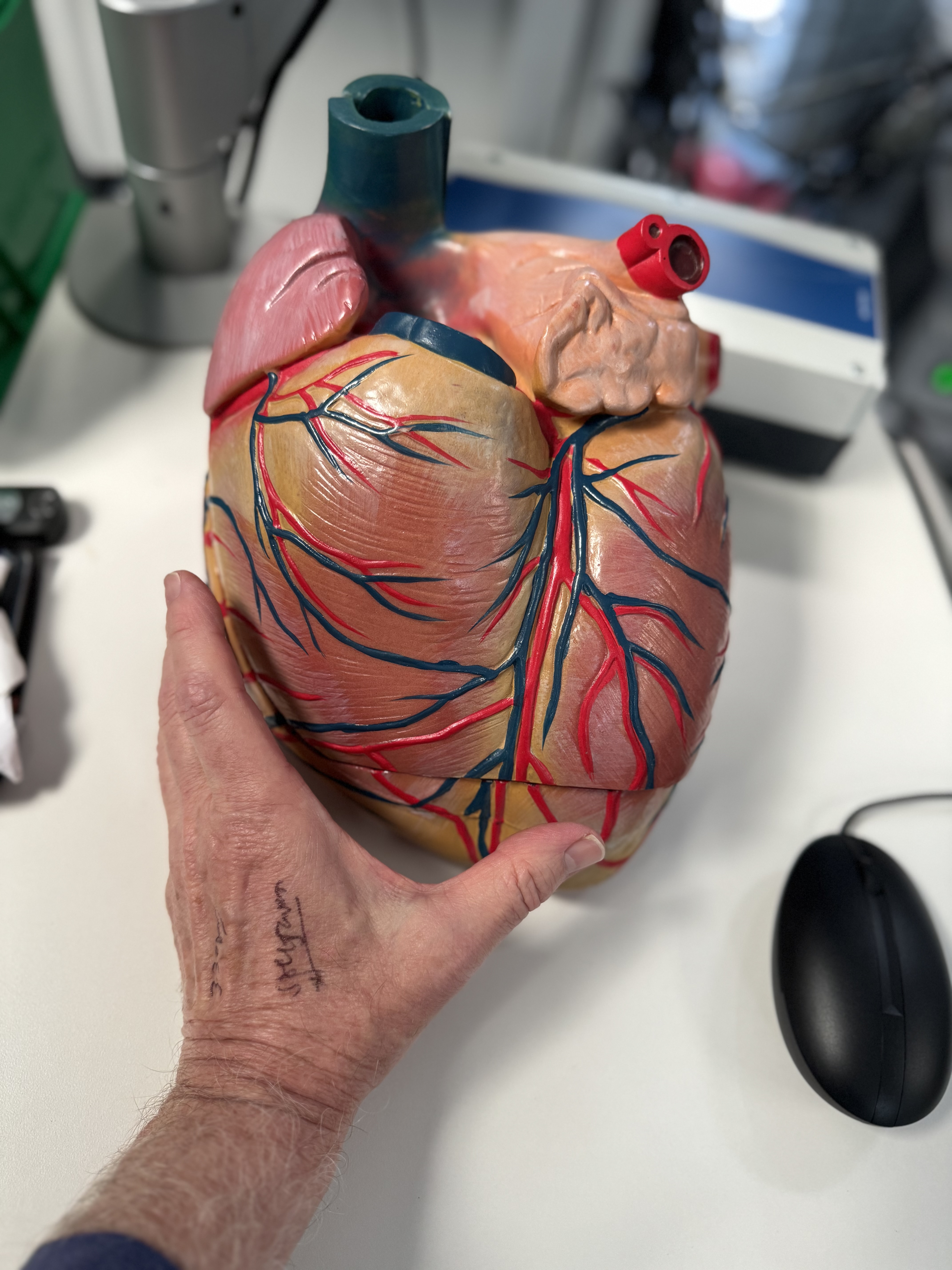
Cause map

The main causes, in plain English.

Etiologic differential, grouped for bedside triage.

01

Coronary artery disease and heart attack

Blocked heart arteries can starve the heart muscle of oxygen. A heart attack can leave scar tissue that does not squeeze well. Sometimes repeated smaller artery problems weaken the heart over time.

Clue: chest pressure, jaw or arm pain, sweating, nausea, or breathlessness can be heart attack symptoms.

Ischemic cardiomyopathy remains a leading cause of HFrEF. Assess CAD probability, ACS, prior infarct, viability where relevant, scar burden, revascularization options, antithrombotic needs, MR from ischemic remodeling, and competing HFpEF/CMD phenotypes.

02

High blood pressure

Long-term high blood pressure makes the heart pump against extra force. The muscle can become thick, stiff, enlarged, or eventually weak.

Pattern: this often contributes to heart failure with normal EF, especially with diabetes, kidney disease, sleep apnea, or atrial fibrillation.

Hypertensive heart disease may drive LVH, LA enlargement, diastolic dysfunction, CKD interaction, pulmonary venous hypertension, and later systolic dysfunction. Consider secondary hypertension when severity, age, potassium, OSA, renal disease, or resistant pattern suggests it.

03

Valve disease

Heart valves are one-way doors. If a valve is too tight or leaks badly, the heart may work too hard, stretch, or face pressure it cannot handle.

Examples: aortic stenosis, mitral regurgitation, mitral stenosis, tricuspid regurgitation, and infection of a valve.

Quantify valve lesion severity, chamber response, pulmonary pressures, rhythm, LV/RV function, symptoms, and intervention timing. Acute severe MR/AR, prosthetic dysfunction, and endocarditis can present as abrupt pulmonary edema or shock.

04

Cardiomyopathy and myocarditis

Cardiomyopathy means a heart muscle disease. Some types are inherited. Others follow viral inflammation, pregnancy, chemotherapy, alcohol, stimulant drugs, thyroid disease, or unknown triggers.

Important: some cardiomyopathies improve when the cause is found and treated early.

Differentiate dilated, hypertrophic, restrictive, arrhythmogenic, stress-induced, peripartum, inflammatory, toxic, genetic, and infiltrative cardiomyopathies. Use CMR, genetic testing, coronary evaluation, EMB, and family screening selectively.

05

Rhythm problems

A heart rhythm that is too fast, too slow, or very irregular can weaken the heart or make symptoms worse. Atrial fibrillation is a common partner with heart failure.

Clue: palpitations, racing pulse, dizziness, fatigue, or breathlessness may point to rhythm trouble.

Evaluate AF/flutter, high PVC burden, SVT, ventricular tachycardia, bradyarrhythmias, pacing-induced cardiomyopathy, chronotropic incompetence, and tachycardia-mediated cardiomyopathy. Rhythm control may be disease-modifying in selected HF patients.

06

Lung pressure and right heart strain

Lung disease, blood clots in the lungs, sleep apnea, or high pressure in the lung arteries can strain the right side of the heart.

Pattern: leg swelling, belly swelling, liver tenderness, and prominent neck veins can be right-sided fluid backup.

Right HF workup should distinguish group 2 PH from pulmonary arterial hypertension, CTEPH, hypoxic lung disease, RV infarct, congenital shunt, severe TR, and mixed phenotypes. Echo estimates are screening, not definitive hemodynamics.

07

Metabolic, endocrine, kidney, and high-output states

Diabetes, obesity, kidney disease, anemia, thyroid disease, severe infection, and some vitamin deficiencies can contribute to heart failure or make it harder to control.

High-output heart failure: rarely, the heart pumps a lot of blood but still cannot keep up with the body's unusually high demand.

Assess CKD/cardiorenal syndrome, diabetes, obesity, iron deficiency, thyroid disease, anemia, AV fistula, cirrhosis, sepsis, beriberi, Paget disease, pregnancy physiology, and obesity-related high-output HF. Treat systemic drivers alongside cardiac congestion.

08

Medicines, alcohol, toxins, and cancer therapy

Some medicines and substances can weaken the heart or hold onto fluid. Examples include heavy alcohol use, cocaine or amphetamines, some chemotherapy, certain targeted cancer medicines, NSAID pain relievers, and some diabetes or rhythm medicines.

Tip: bring a full medicine and supplement list to appointments.

Review anthracyclines, HER2 agents, proteasome inhibitors, TKIs, immune checkpoint inhibitors, alcohol, cocaine, methamphetamine, anabolic steroids, NSAIDs, TZDs, non-DHP calcium channel blockers in HFrEF, and negative inotrope interactions.

Safety

Symptoms that should not wait.

Heart failure can worsen quickly when fluid backs up into the lungs, a heart attack occurs, rhythm becomes unstable, or blood pressure is dangerously high or low.

The patient-facing symptoms below map to acute pulmonary edema, ACS, cardiogenic shock, malignant arrhythmia, PE, hypertensive emergency, severe hypoxemia, and decompensated right HF. Initial priorities are ABCs, ECG, oxygenation, blood pressure, congestion/perfusion profile, troponin, natriuretic peptides, renal function, electrolytes, and trigger identification.

Severe breathlessnessEspecially at rest, lying flat, or with pink frothy sputum.
Chest pain or pressurePossible heart attack or unstable coronary syndrome.
Fainting or confusionPossible low flow, rhythm problem, or severe hypoxemia.
Blue lips or low oxygenPossible pulmonary edema, lung clot, or respiratory failure.
Rapid swelling or weight gainFluid can accumulate before breathing becomes dangerous.

Workup

What doctors usually check.

Testing should confirm HF, phenotype it, find the cause, and identify reversible triggers.

Exam and history

Doctors look for swelling, lung crackles, neck vein pressure, blood pressure, rhythm, weight change, medicines, and possible triggers.

Blood tests

BNP or NT-proBNP can support the diagnosis. Kidney function, electrolytes, anemia, thyroid, diabetes, and heart injury tests may guide treatment.

ECG and rhythm

An ECG checks electrical patterns, old heart attack clues, atrial fibrillation, and dangerous rhythm problems.

Echo and imaging

An echocardiogram estimates EF, valve function, chamber size, pressure clues, and right heart function. Chest X-ray may show fluid or an enlarged heart.

Initial classification

Determine acute versus chronic, de novo versus decompensated, wet/dry and warm/cold profile, LVEF group, RV involvement, shock features, and ICU threshold.

Core tests

ECG, CXR, natriuretic peptide, troponin trend, CMP, CBC, magnesium, TSH, iron studies, HbA1c/lipids, urinalysis/UACR when relevant, and medication/toxin review.

Cardiac imaging

TTE for LVEF, LV geometry, diastology, LA size, RV function, valve disease, pulmonary pressure estimate, IVC, pericardium. Use CMR, TEE, CT, nuclear, or invasive testing by phenotype.

Etiology and triggers

Assess ischemia, valvular disease, AF/PVC burden, myocarditis, infiltrative disease, genetic cardiomyopathy, PH/CTEPH, sleep apnea, renal disease, infection, PE, anemia, and therapy-limiting comorbidities.

Medical visuals

What heart failure can look like.

Chest X-ray showing acute pulmonary edema with enlarged heart and pleural effusions.
Chest X-ray: pulmonary edema. Acute pulmonary edema with enlarged heart size and small pleural effusions. Image by James Heilman, MD, CC BY-SA 3.0.
Lower leg and foot swelling consistent with peripheral edema.
Leg edema. Peripheral swelling is common in right-sided congestion, venous disease, kidney disease, medications, and other causes. Image by Ryaninuk, CC BY-SA 4.0.
Echo: dilated cardiomyopathy. A short echocardiogram clip showing severe mitral regurgitation in dilated cardiomyopathy, an example of poor systolic function. CardioNetworks ECHOpedia, CC BY-SA 3.0.